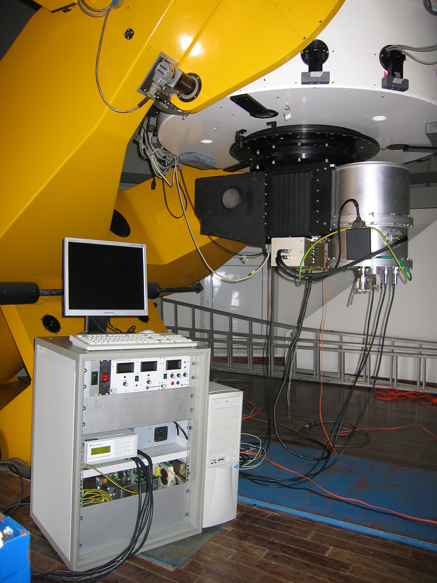
Under a contract with the Foundation for Research and Technology Hellas (FORTH) a camera for the near infrared spectral region (900 nm – 2500 nm) was developed and built by Fraunhofer IOF. Since May 2006 the camera has been working at the 1.3 m telescope of the University of Crete on the Skinakas summit.
To minimize image noise as far as possible the whole camera optical system is set up in vacuum and cooled with liquid nitrogen. The corresponding cryostat allows for a camera operation within the typical range of pointing angles used for observations. The cryostat concept was developed at Fraunhofer IOF while the actual design and construction was undertaken by a specialized company.
The camera enables a re-imaging of the telescope image onto the detector using an optical design according to Offner /2/. With this three-mirror design a very effective suppression of stray light may be obtained by placing a cold stop in front of one of the mirrors. Because of space constraints inside the cryostat with its diameter of 400 mm the optical system had to be made very compact. However, using an aspherical mirror, diffraction-limited performance could be reached even for this compact design. Characteristic parameters of the optical system are:
- Aperture: f/7.7
- Image size: 20 x 20 mm²
- Length of the imaging system: 220 mm
- OPD < λ/4 at 1 μm
To preserve the adjustment of the optics during cool-down, an athermal design with metal mirrors was chosen. These mirrors were fabricated at Fraunhofer IOF by ultra-precision diamond turning. The surfaces obtain a precision of form better than 100 nm and a surface roughness better than 5 nm. To select interesting ranges of wavelength a filter wheel with 8 filters as well as the corresponding driver electronics and software were developed and built.
The whole optical setup was mounted on the cold plate of the cryostat. Prior to integration the positions of the reference points of all optical elements were measured with a precision of < 10 μm. The adjustment of the optical setup relied on gage plates with another set of reference points which were trimmed to a precision of 10 μm according to the measured dimensions of the optical elements.
After integration and adjustment of the optical system the detector chip /2/ and the corresponding driver electronics were mounted. Finally the camera system was characterized and calibrated.
References:
/1/ Offner, A.: Unit Power Imaging Catoptric Anastigmat, United States Patent
3,748,015, July 24, 1973.
/2/ 1 k x 1 k Hawaii chip, Rockwell Science Center, http://www.rsc.rockwell.com/imaging/hawaii1rg.html.
