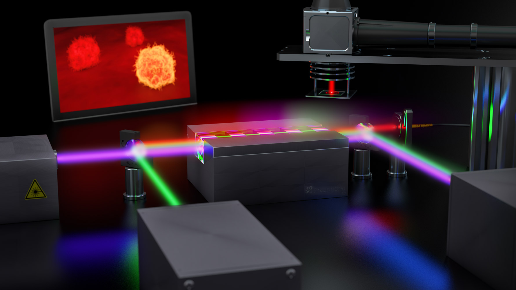
While optical analysis techniques such as microscopy and spectroscopy are already extremely efficient in the visible wavelength range, they quickly reach their limits in the extreme wavelength ranges such as ultraviolet, infrared, or terahertz. A team of researchers at the Fraunhofer Institute for Applied Optics and Precision Engineering IOF took up this challenge and developed a promising quantum imaging solution that can facilitate highly detailed insights into tissue samples using extreme spectral ranges and less light.
Quantum Imaging - making the invisible visible
The operating principle
To make relevant information visible in extreme wavelength ranges, the Jena researchers are using the quantum mechanical effect of photon entanglement. In an inter-ferometric setup, a laser beam is sent through a nonlinear crystal in which it generates two entangled light beams. Even though both beams have very different wavelengths - depending on the crystal’s properties - they are still connected to each other due to their entanglement. So now, while one photon beam in the invisible spectralrange is sent to the object for illumination and interaction, its twin beam in the visible spectrum is captured by a camera. “Since the entangled light particles carry the same information, an image is generated even though the light that reaches the camera never interacted with the actual object," explains quantum researcher Dr. Markus Gräfe of Fraunhofer IOF.
Application in the medical field
One possible field of application that could benefit from this innovative process is medicine. For example, the quantum imaging solution allows previously hidden bio-substances to be distinguished based on their characteristic molecular vibrations. The method promises great progress in the detection and treatment of some types of cancer by visualizing the characteristic concentration or expression of certain proteins. The method can also be used in drug research to gain extensive knowledge about the distribution of bio-substances.
Further technical developments
The research results are obtained within the Fraunhofer Lighthouse Project QUILT. There, besides Fraunhofer IOF also Fraunhofer IPM, Fraunhofer IMS, Fraunhofer ITWM, Fraunhofer IOSB, and Fraunhofer ILT participate. The team is currently working on shrinking the system even more compactly to the size of a shoebox and also further enhance its resolution. To lowering the hurdles for industry users, the long-term goal is to integrate quantum imaging as a basic technology into existing microscopy systems.
